Transforming 3D Printers into PCR Machines for Laboratory Automation
Welcome to an innovative exploration of 3D printing technology in laboratory automation. This page showcases how LabBot, a modified 3D printer, can be configured as a PCR machine, revolutionizing accessibility and sustainability in healthcare tools.
Our system integrates advanced components including a milled thermoblock, an illuminated tube holder for fluorescence imaging, and an Arduino microcontroller shield for precise temperature control. Coupled with Python-based software for thermocycling and imaging, reusable PCR tubes, and a custom pipettor module, LabBot offers a comprehensive solution for real-time monitoring of tube-based reactions using Taqman probe PCR.
Join us as we delve into each step of this groundbreaking process, from mineral oil aspiration to final data analysis, demonstrating the potential of 3D printers in advancing laboratory automation and PCR technology.
Robotic scheduler software for TaqMan probe PCR layout
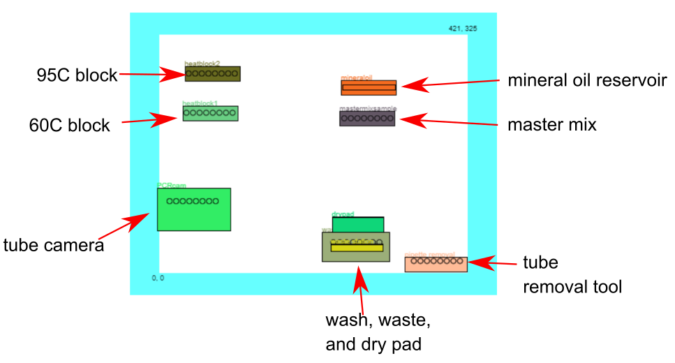
The layout of LabBot deck that illustrates the objects used in the example Taqman probe PCR based nucleic acid detection assay. The LabBot Robotic Schedular software includes this tool where you can define objects to document your method.
Macro for mineral oil aspiration
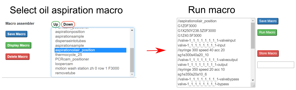
Setting aspiration of mineral oil macro using the LabBot Robotic Scheduling Software
Step 1 Mineral oil aspiration
The LabBot Robotic Scheduler software makes it easy to assemble macros and save them so that you can point, click and run. Here is how an oil aspiration macro was saved, selected, and run.
Master mix aspiration macro
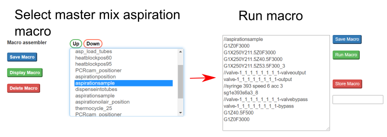
How a macro for sample aspiration can be designed for controlling a 3D printer
Step 2 Master mix aspiration
After the pipettes load the mineral oil, then the master mix is aspirated. They have to be done with these cut down PCR tubes in order to aspirate 10μl of master mix since these pipettes have a gasket membrane that is used for sealing tubes which are done afterward. Aspiration of 8μl of master mix. The tubes contain 10μl.
Step 3 Load tubes using pipette tips
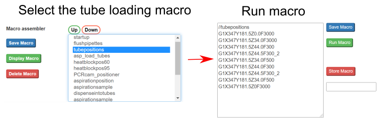
After master mix aspiration the pipette tips are moved to PCR tubes so that they can be loaded and sealed for thermocycling.
Loading tubes so that they can be moved
Step 4 Dispense master mix and mineral oil into tubes
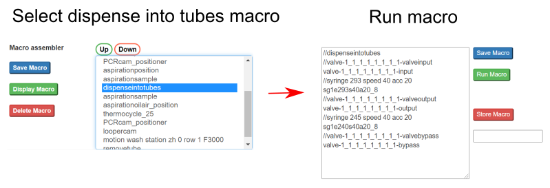
After tube loading, now the pipettes can dispense the master mix and mineral oil that it previously aspirated. Dispense master mix and mineral oil into tubes for thermocycling PCR detection.
Dispensing reaction mix into loaded tubes
Dispensing reaction mix into loaded tubes
Step 5 Thermocycle and imaging reaction
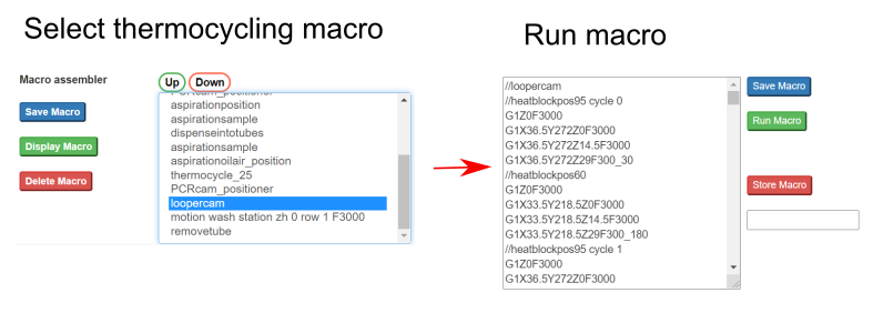
Now the samples and be incubated and it is possible to image after every cycle or every so many cycles. The macro does the imaging after 5 cycles (95C denaturation for 30 seconds and 65C extension for 2 minutes).
Thermocycling by moving the tubes around
Step 6 Remove the tubes
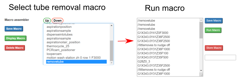
After the reaction is finished, the system includes a technique for removing the tubes. Then the pipettes can be washed and on to the next set of samples. Tube removal technique. After the reaction is finished the tubes are removed using a removal tool
Removal of tubes after the reaction
Example data from a Taqman PCR run on a modified 3d printer
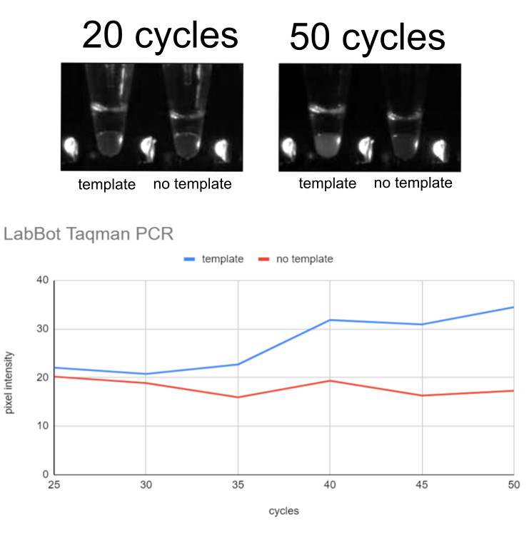
When visualizing the fluorescence (using a blue LED and amber color filter in front of a camera). The Taqman probe oligonucleotide DNA sequence changes color from red to green when it binds to the target sequence. So the approach is to quantify the change of color of the course of the thermocycling process. Comparison between a template and no template is plotted to demonstrate how the PCR amplified nucleic acid signal can be detected using a Taqman probe.